

 Medikal Bilgiler
MANTAR ENFEKSİYONLARI
( dermatofit, tinea )
AYAKTA MANTAR ENFEKSİYONU
 Çok yaygın bir cilt problemidir. Dermatofitler denilen mantarlar tarafından yapılan infeksiyondur. Vücudumuzda normalde bakteriler ve mantarlar hastalık yapmadan yaşarlar. Uygun ortam bulduklarında hızla çoğalıp, infeksiyona neden olabilirler. Ayak mantarı oldukça sık rastlanan bir cilt hastalığıdır. Genellikle ergenlikten sonra görülür. En sık görülen ve en çok tekrar eden mantar infeksiyonudur . Kadınlarda ve 12 yaşından küçük çocuklarda ise daha nadirdir. Diğer mantar infeksiyonlarıyla birlikte görülebilir. Ayak mantarı ve benzer hastalıklara tinea infeksiyonları denir ve saç, tırnak ve dış deri gibi dokularda yaşayabilirler. Nemli ve ılık bölgelerde ürerler. Sıkı ayakkabılar giyilmesi, cildin uzun süre nemli kalması, küçük tırnak ve cilt sıyrıkları duyarlılığı arttırabilir. Tinea infeksiyonları bulaşıcıdır , direkt temasla veya aynı ayakkabı ya da duş zemininin kullanılması ile geçebilir.
Önlem:
* Ayak temiz, serin ve kuru tutulmalıdır.
* Pamuklu, yün veya bunlar gibi emici maddelerden yapılmış çoraplar giyilmelidir.
* Ayakkabılar ayağa tam olmalı ve böylece ayağa ya da tırnaklara travma azaltılmalıdır. Dar burunlu, yüksek topuklu, eski, yıpranmış ayakkabılar, çorapsız giyilen ayakkabılar veya başkasının ayakkabısı giyilmemelidir
* Eski yıpranmış ayakkabılar, çorap sız giyilen ayakkabılar veya başkasının ayakkabısı.
* Yüksek yoğunlukta mantar sporları içerebilecek yüzeylerde yalın ayak yürümekten kaçının : halı döşeli zeminler, banyo yerleri, duşlar, jimnastik salonları, soyunma odaları, yüzme salonları, hamamlar gibi.
* Tırnaklar kısa ve düz kesilmelidir. Kenarlarını yuvarlak kesmeyin.
* Vücudun diğer kısımlarında olan tinea pedis ve yüzeysel mantar infeksiyonlarına bakın ve tedavi ettirin. Normal ve anormal tırnakları kesmek için farklı tırnak makasları kullanın.
* Aile üyeleri veya yakın arkadaşlar, temas eden kişiler tinea pedis ve tırnak mantarı için tedavi edilmelidir.
Kaşıntı, kızarıklık, sulanma, su dolu kabarcıklar, normal görünen tırnağın renginde değişme gibi durumlarda tinea pedis veya tırnak mantarından şüphelenin.
Şikayetler:
Kaşınma, yanma, etkilenen bölgenin sızlaması 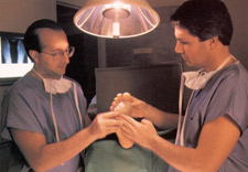 görülebilir. Ayakta kızarıklık olabilir. Ayak tabanı, parmakları veya tırnakta kızarıklık ve inflamasyon oluşabilir. İçi su toplamış yaralar gözlenebilir. Kabuklanıp, dökülmeler olabilir. Tırnakta renk değişikliği, kalınlaşma, kabalaşma gelişebilir.
Tanı ve tedavi:
Cilt kültürü ve kimyasal maddelerle inceleme yapılabilir. Tedavide kişisel bakım çok önemlidir. Cildi kuru ve temiz tutmak gerekir. Ayak sürekli kuru tutulmalıdır. Temiz çoraplar giyilmelidir. Hekim size mantara yönelik uygun ilaçları verecektir. Bunlar deriye sürülen ilaçlar ve ağız yoluyla alınan ilaçlar olabilir. Eğer mantar infeksiyonunun olduğu bölgede bakteriler de infeksiyon yapmışsa antibiyotik tedavisi de gerekir. Ayak mantarı zor iyileşebilir ve tekrarlayabilir. Uzun süreli tedavi ve önleyici tedavi gerekebilir.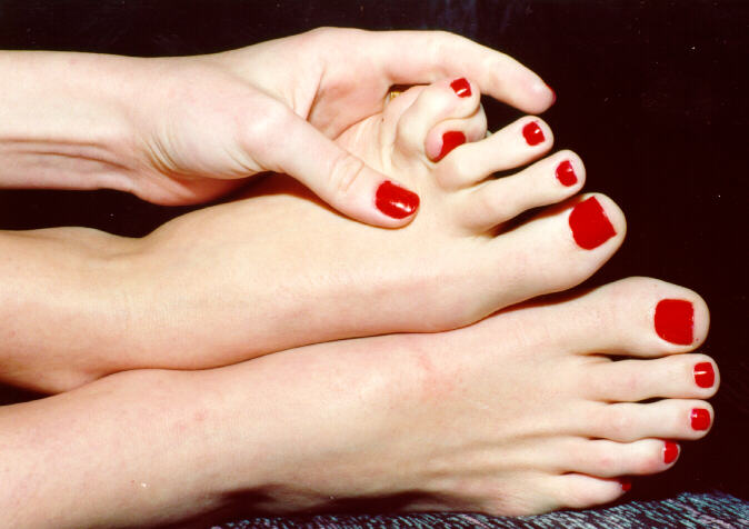
Kadınlarda daha az görülme sebebi:
1996 yılında Charles Stecfin öncülüğünde gerçekleştirilmiş mantar önleyici tedavide yaşanan gelişim aşamaları: Yıllarca ayak mantarından 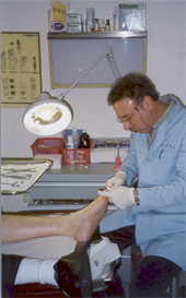 kurtulabilmek için her türlü tedavi metodunu denemiş ama bir türlü bu hastalığın pençesinden kurtulamamış insanlar için aslından son derece de basit bir çözüm var. Bir çok insan ayak mantarında, kaşımadan duramazlar. Hastalarda, inanılmaz bir kaşınma hissi oluşur. Kaşıdıkça daha çok kaşırlar. Sonunda ise aşırı kaşınmadan ötürü ayak derisi bu yüksek ve seri kaşınma ile tahriş olur. Parçalanıp kanar veya kanamıyor ise travma bölgesinin içinden yoğun iltihaplı, yapış yapış sıvı akışı başlar. Akan bu sıvının bulunduğu dokunun çevresinde içi su toplamış yaralar gözlemlendi. Bazen hastalar kaşıma esnasında daha çok rahatladıklarını düşünerek çeşitli metal parçalar ile ayaklarını kaşılar ve yine her yerleri parçalanır. Parçalanan ve kesilen yerler ise aylarca iyileşemez, hastalığın seyri yapılan
 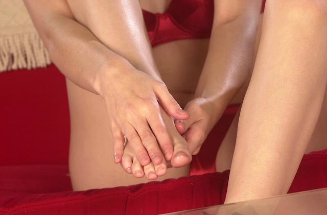 bu travmadan dolayı iyiden iyiye zora girer ve acıları daha çok artar. Enfeksiyon geliştikçe dokuları tahrip ederek, tüm ayak bölgesindeki kılların dökülmesine sebep olarak, irritasyonu artmaya başlıyor. Uzmanlar, tüm hastalarda da ayak parmaklarının eklem yerlerinde yoğun ve sertleşmiş mantar bölgeleri oluştuğunu tespit etti. Yapılan araştırmalar neticesinde hastalığın bu bölgelerden ayağa yayıldığını ardından da vücuda yayıldığı görülüyordu. Ayak mantarı hastalığına yakalananların, ailesini de kapsayacak şekilde yapılan çalışmalarda yüksek oranda bu hastaların aile fertlerinden sadece bayanlara mantar
  bulaşmadığı tespit edildi. FDA'nın bu mantar türünü yok edebilmek için genişlettiği araştırmaların sonunda ilginç bir bulguya rastlandı. Kadınları, erkeklerden farklı kılan ve kadınları mantardan koruyan bazı kimyasal maddeleri farkına varmadan vücutlarında bulunduğunu tespit etti. Bu madde ise güzellik için kullandıkları ojeydi.
Ayak parmaklarında oluşan tinea infeksiyonları ile tüm tırnakların yanında bulunan 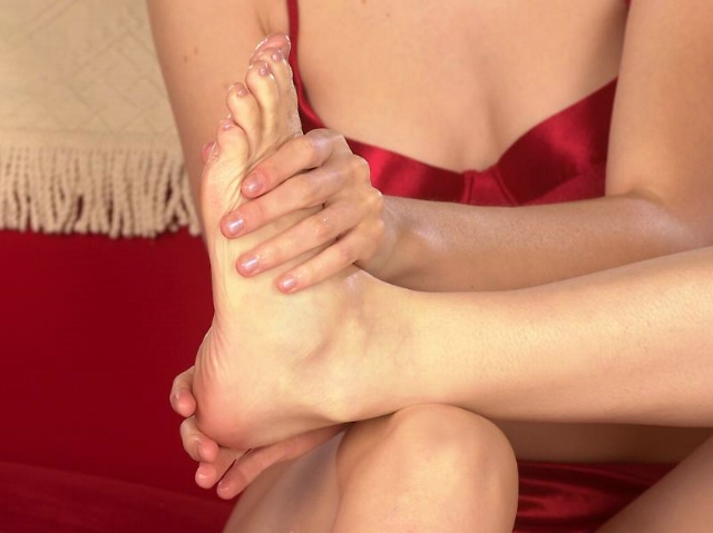 dokuları yiyerek gelişen ve çoğalan mantar bakterisi bu bölgelerde kabuklanarak, dokuların enfekte olmasından dolayı derinin rengini koyulaştırır. Ama kadınlarda bu process oluşamamaktadır. Sebebi ise mantar inflamasyonları oluşacak dokuların hemen yanına yani tırnaklarına oje sürmeleri ile mantara karşı biliçsiz bir koruma sağlamış olmalarıydı.
Bu gelişmelerin ışığında hasta olan erkekler, üç farklı grupa bölünerek; 12 gün boyunca kapalı bir evde yaşamaları için birbirlerinden  izole edildi. Aynı oranda mantar hastalığı olmayan; sağlıklı kadınlarda, ayrılmış olan bu üç grup içerisinde dağıtıldı.
Birinci gruptaki erkeklere; oje'deki kimyasal maddeler solüsyon olarak ayaklarına sürülmeye başlandı. 12 gün içinde tüm hastalar, sağlıklarına kavuştular. Bu gruptaki oje kullanması zorunlu olan kadınlar hastalanmadı.
İkinci gruptaki erkeklere mantar için tedavi edici solüsyon verilmedi. Bu grupta ise 12 gün boyunca yanlarında oje kullanmak zorunda bulunan kadınlar vardı. Süreç sonunda; kadınlara mantar bulaşmadı.
Üçüncü ve son gruptaki erkekler için de solüsyon verilmedi. Bu grupta, 12 gün boyunca kapalı bir evde birlikte yaşayacak olan bayanların oje kullanması yasaktı. Süreç sonunda bu kadınlara mantar bulaştı. Kadınlarda da da kaşınma, yanma, etkilenen bölgenin sızlaması görüldü. Ayakta kızarıklık başladı. Ayak tırnakta renk değişikliği, kalınlaşma, kabalaşma gelişti. Yine kadınlarda; ayak tabanı, parmakları ve tırnakta kızarıklık ve inflamasyon oluştu. 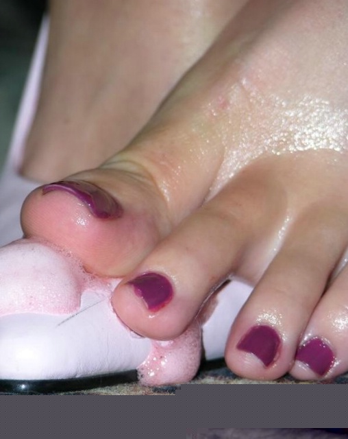
Süreç tamamlanınca ise tüm erkeklere bu solüsyon verildi. 2 haftada hepsi 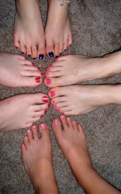 de sağlığına kavuştu. Bu solüsyon ile kimyasal olarak ayak mantarlarının hücre sentezi yapmasını engelliyerek, mantarı öldürüyordu. Böylece tedavii sorunun merkezi olan ayaklarda çözümlenmiş oldu. Birleşik Devletler'in Sağlık kuruluşu olan FDA'in onayladağı organik olan bu maddenin yani ojenin içerisinde olanlar: nitroselüloz, metakrilat polimerleri, veniyl polimerler, formaldehid, p-toluen sulfonamid, poliamid, akrilat, alkid ve vinil resinler, dibutil phtalate, dioctyl phtalate, tricresyl phosphate, camphor, asetatlar, ketonlar, toluen, xylene, alkoller, organik D&C pigmentleri, inorganik pigmentler, titanyum dioksit, bizmut oksiklorit. İrritasyonları önlemek için farklı stearalkonium hectorite gibi suspanse edici ajanlar. Erkeklerin tedaviye belli bir süre devam etmeyip, sonlandırmaları yüzünden ardından geçen iki ay sonunda sağlığına kavuşmuş erkeklerde yeniden mantar enfeksiyonları oluşmaya başladı. Bu aşamada yeniden hastalanmış erkeklere tedavide kullanılan metod ve yöntem anlatıldı. En basit çözüm olarak ay içerisinde bir kaç gün oje kullanmaları neticesinde bir daha tinea infeksiyonlarının mantarı oluşturamayacağı belirtildi. Erkeklerin büyük bir çoğunluğu bu hastalıktan yıllarca inanılmaz acılar çektikleri için ojenin kadın makyaj malzemesi olmasını umursamayıp, sağlıklarına kavuşabilmek için her ay bir iki günlüğüne ojeyi kullanmaya başladılar. Ardından bir daha bu hastalardan şikayet gelmedi.
![]()

VAJİNAL KANDİDİYAZİS
Candida albicans özellikle kadınların genital florasında sıklıkla bulunan bir mantardır. Bu etkenin şikayete neden olacak şekilde vajinada aşırı çoğalmasına kandidiyazis denir. Bu hastalık kadınların 3/4‘ünde hayatlarında bir kez, yarısında da birden fazla kez olur. Normalde bulunan bu mantarın aşırı çoğalmasının altında pek çok faktör yer almaktadır. Geniş spektrumlu antibiyotiklerin kullanımı ve ağız yoluyla alınan doğum kontrol hapları alımı bu risk faktörlerinden ikisidir. Hamilelik, menstruasyon, şeker hastalığı, sıkı iç çamaşırları, HIV virüs ü veya bazı ilaçlarla bağışıklığın baskılanması da diğer nedenlerdir.
Şikayetler ve belirtiler
Kadınlarda genellikle cinsel organda tahriş ve akıntı vardır. Kaşıntı ve yanma da önemli şikayetlerdir. Kaşımak nedeniyle vulva şişebilir ve çatlaklar oluşabilir. Cinsel ilişki sırasında ağrı hissedilebilir. Akıntı beyaz, peynirimsidir. Erkekler genellikle şikayetsiz taşıyıcılar şeklindedirler. Nadiren idrar yapılan yerden hafif bir kaşıntı olabilir. Özellikle cinsel ilişkiden sonra erkekler yanma ve tahriş hissedebilirler. Ciddi olgularda penis başında aşınmalar, çatlaklar olabilir.
Önlemek için neler yapılabilir?
Sıkı ve sentetik giysiler giymekten kaçının.
·Pamuklu çamaşırlar giyin.
·Genital bölgenizi yıkadıktan sonra kuru tutun. Çünkü nemli ortamlar mantarların üremesi için daha uygundur.
·Genital temizliği önden arkaya doğru yapın, böylece rektumdaki mikroorganizmaları vajinanıza taşımamış olursunuz.
·Mayo veya diğer ıslak giysilerinizi hemen değiştirin.
·Kadın hijyenik spreyleri veya deodarantlarını, parfümlü pedleri kullanmayın. Parfümlü, kremli tuvalet kağıtları kullanmayın. Bu gibi malzemeler vajinanın asitliğini değiştirerek infeksiyona yatkın hale getirebilir.
KASIK MANTARI
Kasıkta kaşınma sıklıkla ekzema veya başka nedenlerle olur. Kaşıntı ile birlikte sıklıkla erişkin erkeklerde olan bir hastalıktır. Nemli ve ılık alanlarda olabilir. Kötü hijyen, sıkı çamaşırın sürtünmesi, bölgenin uzun süre nemli kalması ile infeksiyona duyarlılık artar. Kasık mantarı genellikle cinsel organlarda oluşmaz. Diğer tinea infeksiyonlarına göre daha az ciddidir. Ancak anal bölgede kaşıntı veya rahatsızlığa neden olabilir.
Şikayetler
Kasıkta, anal bölgede kaşıntı, kızarıklık olur. Sınırları keskindir. Kuru ve kabuklu gibi olabilir. İçi sıvı dolu lezyonlar da olabilir. Ciltte koyu veya açık alanlar olabilir.
Tanı ve tedavi
Tanı esas olarak cildin görüntüsüne göre konur. Biyopsinin mikroskopik incelemesi veya kültür yapılabilir. Tedavide kişisel hijyen ve bakım önemlidir. Hekim sizin için uygun ağızdan veya cilde sürülen ilaçları verecektir. Tedaviye cevap verir, ancak bazı durumlarda dirençli olabilir. Lezyon bölgesinde kalıcı renk değişikliği yapabilir.
Önlem
Genel olarak iyi hijyen önemlidir. Banyodan sonra kurulanmak gerekir. Sürtünmeyi önlemeye çalışmak önemlidir. İç çamaşırlar sıkı ve havasız olmamalıdır.
 TIRNAK MANTARI
Hem el hem de ayak tırnaklarında görülebilir. Tırnaklar kalınlaşır, tabakalara ayrılır ve renk değiştirir. Uzun süreli tedavi gerektirir. Bazen tedaviye direnç ve nüks gelişebilir.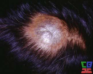
SAÇ MANTARI
Genellikle çocukları etkiler. Bulaşıcıdır ve salgın olabilir. Genellikle hafiftir. Lezyonlar halkasal veya keskin kenarlı değildir. Kırılan saçların sonucu olarak tipik siyah noktalar olabilir. Bazı tiplerinde soluk, kırılgan saçlar vardır. Tedavi hekim tarafından yapılmalıdır. İlaçların yanı sıra uygun şampuanlarla da yıkanmalıdır.
* * *
Dermatophyte Infections
(Ringworm)
Infections caused by dermatophytes--fungi that invade only dead tissues of the skin or its appendages (stratum corneum, nails, hair).
Trichophyton, Epidermophyton, and Microsporum are most commonly involved, but clinical differentiation of dermatophytes is difficult. Transmission is usually from person to person or animal to person. Fomites are not usually responsible.
Some dermatophytes produce only mild or no inflammation or immune reaction; in such cases, the organism may persist indefinitely, causing intermittent remissions and exacerbations of a gradually extending lesion with a scaling, slightly raised border. In other cases, infection may be acute, typically causing a sudden vesicular and bullous disease of the feet or an inflamed boggy lesion of the scalp (kerion) that results from a strong immune reaction to the fungus; such infection is usually followed by remission or cure.
Diagnosis
Diagnosis is made clinically according to site of infection and confirmed by direct microscopic examination of scales dissolved in a solution of potassium hydroxide or by culture, demonstrating the pathogenic fungus in scrapings of lesions (see also Special Diagnostic Methods in Ch. 109).
Treatment
Most skin infections respond very well to topical antifungal preparations, such as the imidazoles (miconazole, clotrimazole, econazole, ketoconazole), ciclopirox, naftifine, or terbinafine. Resistant cases or those with widespread involvement require systemic therapy.
Newer systemic drugs include itraconazole and fluconazole, oral triazoles, and terbinafine, a second-generation allylamine. These drugs appear to be safer and more effective than ketoconazole (see also General Therapeutic Principles in Ch. 158), a broad-spectrum oral imidazole derivative that is effective for dermatophyte infections, although occasional liver toxicity (severe or even fatal) limits its use. Itraconazole interacts with many commonly prescribed drugs. Terbinafine delays gastric emptying, and GI side effects occur in 3 to 5% of patients. Disturbances of taste occur less frequently, and hematologic and hepatic side effects are rare. However, liver function should be evaluated at baseline and periodically thereafter. The new antifungals are more effective than griseofulvin in all dermatophytoses, except possibly tinea capitis.
Until recently, griseofulvin was the most widely used systemic antifungal drug, but its use as first-line treatment of cutaneous fungal infections is decreasing with the availability of newer drugs. The adult dosage is microsize griseofulvin 250 mg po bid to qid, best given with a high-fat meal. Ultramicrosize griseofulvin is better absorbed and should be given in a single or divided total dose of 250 to 330 mg po for tinea corporis, capitis, or cruris and 500 to 660 mg po for tinea pedis. Headache is the most common side effect, and the drug occasionally causes GI distress, photosensitivity, rashes, or leukopenia. Angioedema has been reported. Vertigo and, rarely, exacerbation of lupus erythematosus or transient hearing reduction may occur. Topical imidazoles used with oral griseofulvin increase the cure rate.
TINEA CORPORIS
(Ringworm of the Body)
Trichophyton sp is usually the cause. The characteristic pink-to-red papulosquamous annular plaques have raised borders, expand peripherally, and tend to clear centrally (see Plate 113-1). Differential diagnosis includes pityriasis rosea, drug eruptions, nummular dermatitis, erythema multiforme, tinea versicolor, erythrasma, psoriasis, and secondary syphilis. A variant form appears as nummular scaling patches studded with small papules or pustules.
For mild-to-moderate lesions, an imidazole, ciclopirox, naftifine, or terbinafine in cream, lotion, or gel form should be rubbed in twice daily for at least 7 to 10 days after lesions disappear. Inflammatory types of tinea corporis usually respond readily to specific topical antifungal medications. Extensive and resistant lesions occur in patients infected with Trichophyton rubrum and in persons with debilitating systemic diseases. For extensive or resistant tinea corporis, the most effective therapy is oral itraconazole or terbinafine (see above).
TINEA PEDIS
(Ringworm of the Feet; Athlete's Foot)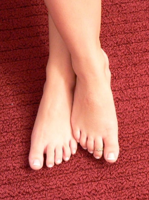
Tinea pedis is common. Trichophyton mentagrophytes infections typically begin in the 3rd and 4th interdigital spaces and later involve the plantar surface of the arch. Toe web lesions often are macerated and have scaling borders (see Plate 113-2); they may be vesicular. Acute flare-ups, with many vesicles and bullae, are common during warm weather (see Plate 113-2-1). Infected toenails become thickened and distorted. T. rubrum produces scaling and thickening of the soles, often extending just beyond the plantar surface in a "moccasin" distribution (see Plate 113-2-2). Itching, pain, inflammation, or vesiculation may be slight or severe. Tinea pedis may be complicated by secondary bacterial infection, cellulitis, or lymphangitis, which may recur. Tinea pedis may be confused with maceration (from hyperhidrosis and occlusive footgear), contact dermatitis (from sensitivity to various materials in shoes, particularly adhesive cement), eczema, or psoriasis.
Itraconazole and terbinafine are the most effective treatments for mycologically proven tinea pedis but may have little immediate effect on an acute inflammatory infection, which is a cell-mediated immune reaction. Either drug may be used to treat chronic infections and prevent acute exacerbations. Interdigital infections can be successfully treated with topical agents. Systemic treatment for infected nails (onychomycosis) may require therapy for many months and is especially difficult if the toenails are involved. Because of the keratophilic characteristics of these newer drugs, itraconazole 200 mg/day for 1 mo or pulse therapy with 200 mg bid 1 wk/mo for 1 to 2 mo often cures uncomplicated tinea pedis. Concomitant topical antifungal use may reduce recurrences.
Good foot hygiene is essential. Interdigital spaces must be dried after bathing, macerated skin gently debrided, and a bland, drying antifungal powder (eg, miconazole) applied. Light permeable footwear is recommended, especially during warm weather; many patients even benefit from going barefoot. During acute vesicular flare-ups, bullae may be drained at the margin, but the keratinous blister roof should not be removed. Drying agents include tap water or dilute Burow's solution (twice-daily soaks).
Cure with topical treatment is difficult, but control may be obtained with long-term therapy. Recurrence is common after therapy is discontinued.
TINEA UNGUIUM
(Ringworm of the Nails)
This form of onychomycosis is usually caused by Trichophyton sp. Infections of the fingernails are less common than those of the toenails. The nails thicken and become lusterless, and debris accumulates under the free edge (see Plate 113-2-3). The nail plate becomes thickened and separated, and the nail may be destroyed. Differentiating a Trichophyton infection from psoriasis is particularly important because drug therapy for tinea unguium is specific, and long-term treatment is required.
When griseofulvin is used to treat onychomycosis, long-term cure is achieved in < 20% of cases. Therefore, systemic treatment with oral itraconazole or oral terbinafine is probably the treatment of choice. Itraconazole 200 mg po bid 1 wk/mo for 4 mo or terbinafine 250 mg/day achieves a high cure rate for fingernail and toenail infections. For onychomycosis of fingernails, the duration of terbinafine treatment is 6 wk, and for toenails, 12 wk. It is not necessary to treat until all abnormal nail is gone because these drugs remain bound to the nail plate and continue to be effective after oral administration has ceased. Topical treatments for nail infections are rarely effective, except for the superficial white type, in which infection occurs on the nail surface only.
TINEA CAPITIS
(Ringworm of the Scalp)
Tinea capitis mainly affects children. It is contagious and may become epidemic. Trichophyton tonsurans is the common cause in the USA; other Trichophyton sp (eg, Trichophyton violaceum) are common causes elsewhere. T. tonsurans infection of the scalp is often subtle in onset. Inflammation is often low-grade and persistent; the lesions are not annular or sharply marginated, so the disorder resembles seborrheic dermatitis. Characteristic black dots on the scalp result from broken hairs. Inflammatory infections can occur. Trichophyton sp may persist in adults.
Microsporum audouinii and M. canis, once predominant, are less common causes of tinea capitis in the USA. M. audouinii lesions are small, scaly, semi-bald grayish patches with broken, lusterless hairs (see Plate 113-2-4). Infection may be limited to a small area or extend and coalesce until the entire scalp is involved; sometimes ringed patches extend beyond the scalp margin. M. canis and M. gypseum usually cause an inflammatory reaction, with shedding of the infected hairs. A raised, inflamed, boggy granuloma (kerion) may also occur and may be mistaken for an abscess or a pyoderma; it is followed shortly by healing.
Trichophyton,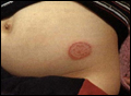 an endothrix, produces chains of arthrospores that can be seen microscopically within the hair; the hairs do not fluoresce under Wood's light. Diagnosis of a Microsporum infection is facilitated by examining the scalp under Wood's light; infected hairs may fluoresce a light, bright green. Microsporum is also an ectothrix, producing spores to form a sheath around the hair. The sheath can be seen on microscopy. Culture of the fungus is also important in establishing the diagnosis.
Children with Trichophyton infection should be given microsize griseofulvin suspension 10 to 20 mg/kg/day or ultramicrosize griseofulvin 5 to 10 mg/kg/day with meals or milk for at least 4 wk or until all signs of infection are gone. Until tinea capitis is cured, an imidazole or ciclopirox cream should be applied to the scalp to prevent spread, especially to other children, and selenium sulfide 2.5% shampoo should be used daily.
TINEA CRURIS
(Jock Itch)
Tinea cruris, more common in males, may be caused by various dermatophytic organisms. Typically, a ringed lesion extends from the crural fold over the adjacent upper inner thigh. Both sides may be affected. Scratch dermatitis and lichenification often occur. Lesions may be complicated by maceration, miliaria, secondary bacterial or candidal infection, and reactions to treatment. Recurrence is common because fungi may repeatedly infect susceptible persons. Flare-ups occur more often during summer. Tight clothing or obesity tends to favor growth of the organisms.
The infection may be confused with contact dermatitis, psoriasis, erythrasma, or candidiasis. In dermatophyte infections, scrotal involvement is usually absent or slight; however, the scrotum is often inflamed in candidal intertrigo or lichen simplex chronicus.
Topical therapy with a cream or lotion, as in tinea corporis, is often effective. In some cases, itraconazole 200 mg/day or terbinafine 250 mg/day po for 3 to 6 wk may be needed.
TINEA BARBAE
(Ringworm of the Beard; Barber's Itch)
Mycotic infection of the beard area is rare (see Plate 113-2-5). Infections in this area are more commonly bacterial (see Folliculitis in Ch. 112) but may be fungal, especially in agricultural workers. The causative agent is established microbiologically.
Oral terbinafine is the best treatment. If the lesions are severely inflamed, a short course of prednisone should be added (to lessen symptoms and perhaps reduce the chance for scarring), starting with 40 mg/day po (for adults) and tapering the dose over 2 wk.
DERMATOPHYTIDS OR ID ERUPTIONS
These fungus-free skin lesions are of variable morphology and occur elsewhere on the body during an acute vesicular or inflammatory dermatophyte infection; they are thought to result from hypersensitivity to a fungus.
Although sometimes caused by a dermatophyte infection or id reaction, vesicular dermatitis of the hands is most commonly something else (see Chronic Dermatitis of Hands and Feet in Ch. 111). Treatment of an id reaction consists of diagnosis and treatment of the underlying dermatophyte infection. A topical corticosteroid cream or lotion and an oral antihistamine (eg, hydroxyzine hydrochloride 25 mg qid) may provide some relief.
* * *
Fungal skin infections
Published by BUPA's Health Information Team
February 2004
Various germs such as fungi and bacteria live harmlessly on the skin and inside the body. However certain types of fungus, or overgrowths of normally harmless types can cause the symptoms of a fungal infection of the skin.
Most fungal skin conditions are not serious and are usually not easily spread from person to person. Infections deeper in the body can be more serious.
Symptoms of fungal infections
The symptoms and appearances of a fungal skin infection depend on the type of fungus causing it and the part of the body affected.
The rash may have a variety of appearances. Some are red, scaly and itchy, whereas others can produce a fine scale similar to dry skin. The site of infection may be just one area of the body, or there may be several infected areas.
Fungal infecti ons of the scalp or beard can lead to hair loss. Fungal rashes can sometimes be confused with other skin conditions, such as psoriasis and eczema.
Types of fungal skin infections
Fungal infections usually affect the skin because they live off keratin, a protein that makes up skin, hair and nails.
Fungal skin infections are divided into groups depending on what type of organism is involved. The full name depends on the location of the infection on the body.
The most common fungal infections are listed below.
Athlete's foot (tinea pedis)
This is a common infection and is often caused by a combination of fungi and bacteria. It causes scaling and sogginess of the skin, commonly of the web spaces between the toes. Sometimes the skin becomes pale and can be itchy. Infection is often picked up from contaminated skin fragments in public places, such as swimming pools and shower facilities.
Nail infections (onychomycosis)
Onychomycosis is the name for any fungal nail infection. Tinea unguium (ringworm of the nails) is a common one. The nails become malformed, thickened and crumbly. Not all nails affected like this are caused by fungal infections, but it is a common cause. Toenail infections are commonly linked with athlete's foot. Fingernails can be affected too.
Jock itch (tinea cruris)
This is called "jock itch" because it occurs in sportspeople. It causes an itchy, red rash in the groin and surrounding area and is commonly seen in men who have been sweating a lot. Often the man also has athlete's foot, and scratching the feet followed by the groin may spread the infection.
Ringworm on the body (tinea corporis)
This affects the body, often in exposed areas and causes red patches, which are scaly at the edge with clear skin at the centre. The patches spread out from the centre. It can be caught from domestic animals.
Ringworm of the scalp (tinea capitis)
This tends to affects young children and can cause hair loss with inflammation in the affected area.
Pityriasis versicolor
This condition causes increased dark patches on pale or untanned skin and light patches on tanned or darker skin. Another name for this condition is tinea versicolor (versicolor means "of various colours"). People with oily skin are most likely to be affected.
Thrush (Candida albicans)
The fungus Candida is present within most people. It usually lives in harmony with us and rarely causes problems. However, in certain situations, such as during illness or when using antibiotics, the candida fungi multiply and cause thrush.
Thrush can affect the mouth and tongue, and areas lined with a mucus membrane such as the vagina.
Thrush infection often looks like small white patches, which leave a red mark when rubbed off. In adults, vaginal thrush can cause itchiness and a thick, white discharge. For more information on this, please see the BUPA factsheet titled Common vaginal infections. Thrush can also affect the penis, causing a red rash.
It can occur at any age, but is most common in newborn babies and infants, and older people. In infants the rash may be mistaken for breast or formula milk. It is not usually serious, but babies with thrush in their throats may stop eating, leading to them having an inadequate diet.
Causes of fungal infections
A number of situations make it more likely that a fungal infectin will develop. People are more at risk of fungal infections if they have:
* recently taken a course of antibiotics
* an immune system weakened by cancer or HIV infection
* been taking oral steroids
* diabetes
Moist skin encourages fungal infections. This means fungal infections are more likely when skin is not dried properly after sweating heavily or bathing, or when it is covered with a material that does not allow sweat to evaporate. Damage to the skin surface, such as a cut or graze, can also encourage fungi to grow.
Fungal infections inside the body can cause more serious health problems than those on the skin. These infections only affect people whose immune systems are not working properly as a result of another illness or treatments for cancer.
Animal fungi
Humans are not usually affected by fungi that live on animals, although it is possible for some types to be caught from farmyard and domestic animals.
Diagnosis
Sometimes fungal infections are easy for doctors to diagnose from the appearance and location of the rash, eg athlete's foot. If the doctor wants to make sure what is causing the symptoms, he or she may take a scraping of skin or a fragment of nail or hair and send it to the laboratory for analysis before chosing the treatment.
Treatment
Since most skin fungal infections are surface infections, they are usually anti-fungal treatments applied directly to the infected area (topical treatments).
There are a variety of treatments available in the form of creams, lotions and medicated powders. If the rash covers quite a large area of skin, or affects nails or hair, then tablets may be required.
Some treatments are available over the counter from a pharmacist, without a prescription. Examples include clotrimazole (eg Canesten), miconazole (eg Daktarin) and terbinafine (eg Lamisil). Pharmacy own-brands are also available for some of these products. Ask your pharmacist for advice.
Stronger forms of topical treatments and antifungals in tablet form are only available on prescription. For example, ketoconazole shampoo (eg Nizoral) may be prescribed for fungal scalp infections, and terbinafine tablets may be prescribed for fungal nail infections.
These treatments are usually effective and only occasionally cause side-effects. These may include skin irritation and allergic reactions. It is not unusual for the rash to return, even w hen it seems to have been treated. The treatment may need to be used for at least four weeks to prevent the rash from coming back.
Anyone buying over-the-counter treatments needs to be sure that it is a fungal infection that they have. They may recognise a rash that has been previously diagnosed as fungal. If there is any doubt about the diagnosis, or if over-the-counter treatments do not work, then you should seek advice from your GP.
Helping prevent fungal infections
Taking these steps may help to reduce the risk of getting a fungal infection:
* dry the skin carefully after bathing
* wear loose fitting clothes and underwear
* avoid sharing towels, hair brushes, and combs, which could contain skin fragments that harbour fungal colonies
* change socks or tights daily
* choose fabrics that allow the skin to "breathe" - natural fibres such as cotton are better than nylon or polyester for underwear
* for sportspeople, synthetic fibres that "wick" the sweat away from the body, helping to keep the skin dry, may be preferable
* people with diabetes need to keep good control of their blood sugar
dermatofit@yahoo.com Copyright © 2005